
The retina is an essential part of the eye responsible for transmitting visual information to the brain. As such, any disorder affecting the retina can result in vision loss or impairment. In this article, we will provide an overview of some of the most common retinal disorders, such as age-related macular degeneration, diabetic retinopathy, and retinal detachment.
Understanding the Retina and Its Function
The retina is a multi-layered tissue located at the back of the eye. Its primary function is to convert light into neural signals that are then transmitted to the brain for interpretation. The retina consists of various types of cells, including photoreceptors, which are responsible for detecting light, and ganglion cells, which transmit visual information to the brain.
The retina is a complex structure with many important components. One of the most important components of the retina is the fovea, which is a small, central area of the retina that is responsible for sharp, detailed vision. The fovea contains a high concentration of cones, which are responsible for detecting color and high levels of light. This is why the fovea is so important for tasks that require visual acuity, such as reading or driving.
Anatomy of the Retina
The retina can be divided into several distinct layers, each with a specific function. The outermost layer, known as the pigment epithelium, absorbs excess light and provides nourishment to the retina. The innermost layer, the ganglion cell layer, contains the ganglion cells responsible for transmitting visual information to the brain.
The retina also contains several other layers, including the outer nuclear layer, which contains the cell bodies of the photoreceptors, and the inner nuclear layer, which contains the cell bodies of other types of cells, including bipolar cells and horizontal cells. These cells play important roles in processing visual information before it is transmitted to the ganglion cells. Click here to read more about What Does Healthy Eating Mean?
The photoreceptor layer, located between the pigment epithelium and the ganglion cell layer, contains two types of cells called rods and cones. Rods are responsible for detecting low levels of light, while cones are responsible for detecting color and high levels of light. There are three types of cones, each of which is most sensitive to a different range of wavelengths of light. This allows us to see a wide range of colors.
Glaucoma is a serious eye condition that can cause gradual vision loss if left untreated. While the symptoms of glaucoma may vary, they often develop slowly and can go unnoticed until the condition has progressed. One common Glaucoma symptom is the gradual loss of peripheral vision. Individuals with glaucoma may also experience blurred vision, difficulty adjusting to low-light conditions, and increased sensitivity to glare. As the disease progresses, the optic nerve can become damaged, leading to permanent vision loss. Regular eye exams are crucial in detecting glaucoma early and preventing further damage.
The Role of the Retina in Vision
The retina is a vital component of the visual system, serving as the primary site for visual processing. Visual information is transmitted from the retina to the brain, where it is interpreted to form the images we see. As such, any disorder affecting the retina can result in vision loss or impairment.
There are many different disorders that can affect the retina, including age-related macular degeneration, diabetic retinopathy, and retinal detachment. These disorders can cause a range of symptoms, including blurry vision, blind spots, and even complete vision loss.
Fortunately, there are many treatments available for retinal disorders, including medications, laser therapy, and surgery. With proper treatment, many people with retinal disorders are able to maintain their vision and lead full, active lives.
Age-Related Macular Degeneration (AMD)
Age-related macular degeneration (AMD) is a progressive eye disease that affects the macula, a small area in the center of the retina responsible for sharp vision. The macula is responsible for our ability to read, recognize faces, and see fine details. AMD is the leading cause of vision loss among people over the age of 50.
AMD is a complex disease that can have a significant impact on an individual’s quality of life. It can make it difficult to perform daily tasks, such as driving, reading, and watching television. In severe cases, it can lead to legal blindness.
Types of AMD
AMD can be classified into two broad categories: dry AMD and wet AMD. Dry AMD is the most common type, accounting for around 85% of all cases. It is characterized by the buildup of drusen, yellow deposits beneath the retina, that can lead to the degeneration of the macula. Dry AMD progresses slowly and can take years to develop.
Wet AMD, on the other hand, is a less common but more severe form of the disease. It is characterized by the growth of abnormal blood vessels beneath the retina, which can leak fluid and blood, causing scarring and damage to the macula. Wet AMD can cause rapid and severe vision loss if left untreated.
Symptoms and Diagnosis
The early stages of AMD may not cause any noticeable symptoms. As the disease progresses, however, individuals may experience blurred or distorted central vision, difficulty reading, and difficulty recognizing faces. AMD can be diagnosed through various methods, such as a comprehensive eye exam, retinal imaging, and visual acuity testing. Early detection and treatment are critical for preserving vision and preventing further damage.
Treatment and Prevention
Currently, there is no known cure for AMD. However, certain treatments may help slow the progression of the disease and preserve vision. These treatments include anti-VEGF therapy, which involves injections into the eye to decrease the growth of abnormal blood vessels, and photodynamic therapy, which uses a light-activated drug to destroy abnormal blood vessels.
Preventing AMD is essential for maintaining healthy eyesight. Individuals are advised to maintain a healthy lifestyle, eat a nutritious diet, avoid smoking, and protect their eyes from UV radiation. Regular eye exams are also essential for early detection and treatment of AMD.
In conclusion, AMD is a complex and progressive eye disease that can have a significant impact on an individual’s quality of life. Understanding the types, symptoms, diagnosis, treatment, and prevention of AMD is essential for maintaining healthy eyesight and preventing vision loss.
Diabetic Retinopathy
Diabetic retinopathy is a serious complication of diabetes that affects the eyes. It occurs when high levels of blood sugar damage the blood vessels in the retina, which is the part of the eye that senses light and sends signals to the brain. Over time, this damage can lead to vision loss and blindness. In fact, diabetic retinopathy is one of the leading causes of blindness among working-age adults.
There are two stages of diabetic retinopathy: non-proliferative diabetic retinopathy and proliferative diabetic retinopathy. In the early stages of the disease, non-proliferative diabetic retinopathy, blood vessels in the retina may become blocked or leaky, resulting in the formation of microaneurysms and swelling in the macula. This can cause mild to moderate vision problems, such as blurred or distorted vision.
In proliferative diabetic retinopathy, new blood vessels begin to grow in the retina, which can lead to scarring and retinal detachment. This can cause severe vision loss and even blindness. If you have diabetes, it is important to have regular eye exams to detect diabetic retinopathy early, when it is most treatable.
Risk Factors for Diabetic Retinopathy
Individuals with diabetes are at an increased risk of developing diabetic retinopathy, particularly if their blood sugar is poorly controlled, they have high blood pressure, or they have had diabetes for a long time. Other risk factors for diabetic retinopathy include smoking, high cholesterol levels, and pregnancy.
If you have diabetes, it is important to manage your blood sugar levels and blood pressure to reduce your risk of developing diabetic retinopathy. You should also quit smoking, if you smoke, and have regular eye exams to monitor for changes in the retina.
Symptoms of Diabetic Retinopathy
Early stages of diabetic retinopathy may not have any noticeable symptoms. As the disease progresses, however, you may experience symptoms such as:
- Blurred or distorted vision
- Floaters, which are small specks or spots that float across your field of vision
- Dark or empty spots in the center of your vision
- Poor night vision
- Colors appearing faded or washed out
If you experience any of these symptoms, it is important to see an eye doctor right away.
Treatment Options for Diabetic Retinopathy
Treatment for diabetic retinopathy depends on the severity of the disease. In the early stages, your doctor may recommend monitoring your condition and making lifestyle changes to manage your diabetes and blood pressure. This may include eating a healthy diet, exercising regularly, and taking medication to control your blood sugar and blood pressure.
If your diabetic retinopathy is more advanced, your doctor may recommend laser therapy. This involves using a laser to seal leaking blood vessels and prevent the growth of new ones. Another treatment option is anti-VEGF therapy, which involves injecting medication into the eye to reduce swelling and inflammation.
If you have diabetic retinopathy, it is important to work closely with your doctor to manage your condition and prevent vision loss. By monitoring your blood sugar and blood pressure, attending regular eye exams, and following your doctor’s treatment plan, you can help protect your vision and maintain your overall health.
Retinal Detachment
Retinal detachment is a serious eye condition that occurs when the retina becomes detached from the underlying tissue responsible for providing it with essential nutrients and oxygen. If left untreated, retinal detachment can lead to permanent vision loss.
The retina is a thin layer of tissue located at the back of the eye that is responsible for detecting light and transmitting signals to the brain. When the retina becomes detached, it can no longer function properly, which can result in vision loss or blindness.
Causes and Types of Retinal Detachment
Retinal detachment can be caused by various factors, such as trauma to the eye, inflammation, or structural abnormalities in the retina. Individuals who are nearsighted, have a family history of retinal detachment, or have had cataract surgery are at a higher risk for developing retinal detachment.
There are three types of retinal detachment: rhegmatogenous retinal detachment, tractional retinal detachment, and exudative retinal detachment. Rhegmatogenous retinal detachment occurs when a tear or hole develops in the retina, allowing fluid to leak in and separate the retina from its supporting tissue. Tractional retinal detachment occurs when scar tissue on the retina pulls it away from the underlying tissue. Exudative retinal detachment occurs when fluid builds up beneath the retina due to inflammation or a growth such as a tumor.
Symptoms and Diagnosis
The symptoms of retinal detachment may include sudden flashes or floaters in the vision, a shadow or veil covering part of the visual field, or a sudden decrease in vision. It is important to seek medical attention immediately if you experience any of these symptoms, as early diagnosis and treatment can help prevent permanent vision loss.
Retinal detachment can be diagnosed through various methods, such as a dilated eye exam, ultrasound imaging, or optical coherence tomography. During a dilated eye exam, an eye doctor will examine the retina using a special instrument and may use eye drops to dilate the pupils for a better view of the retina.
Surgical Treatments and Recovery
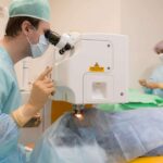
The standard treatment for retinal detachment is surgery, which may include laser therapy, cryotherapy, or scleral buckling, depending on the type and severity of the detachment. Laser therapy involves using a laser to seal the tear or hole in the retina, while cryotherapy involves using freezing temperatures to create a scar that seals the tear or hole. Scleral buckling involves placing a band around the eye to push the wall of the eye against the retina, helping it to re-attach.
Following surgery, individuals will need to attend regular follow-up appointments to monitor their progress and ensure that the retina has re-attached to the underlying tissue. Recovery time may vary depending on individual circumstances, but most individuals can expect to resume normal activities within a few weeks to a few months.
In conclusion, retinal detachment is a serious eye condition that requires prompt medical attention. Early diagnosis and treatment can help prevent permanent vision loss and improve the chances of a successful recovery.
Conclusion
Retinal disorders can significantly impact an individual’s quality of life. Age-related macular degeneration, diabetic retinopathy, and retinal detachment are just a few examples of the numerous conditions that can affect the retina. However, with proper management and treatment, individuals with retinal disorders can often maintain adequate vision and prevent further vision loss.